38 sperm cell diagram with labels
Specialised animal cells - Cell structure - BBC Bitesize Sperm The head contains the genetic material for fertilisation in a haploid nucleus. The acrosome in the head contains enzymes so that a sperm can penetrate an egg. The middle piece is packed with... Diagram Of A Sperm Cell Illustrations, Royalty-Free Vector ... - iStock Browse 420 diagram of a sperm cell stock illustrations and vector graphics available royalty-free, or start a new search to explore more great stock images and vector art. Newest results structure of a sperm cell Education Chart of Biology for Reproduction Process of Human... Cell potency. From Totipotent to Pluripotent, Multipotent, and...
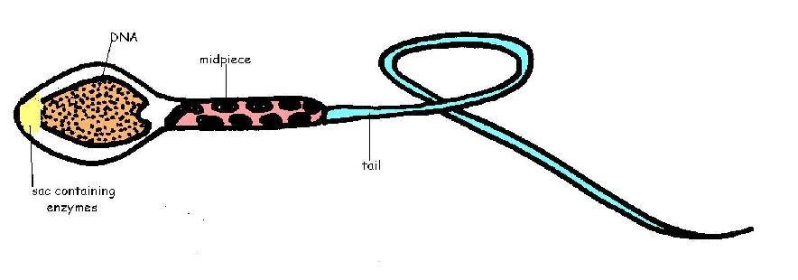
labelled diagrams - the sperm cell labelled diagrams - the sperm cell to the right is a detailed 2D diagram of the sperm cell. there are many parts of a sperm cell. it is extremely small compared to the female egg.

Sperm cell diagram with labels
Sperm - Wikipedia The mammalian sperm cell can be divided in 2 parts: Head: contains the nucleus with densely coiled chromatin fibers, surrounded anteriorly by a thin, flattened sac called the acrosome, which contains enzymes used for penetrating the female egg.It also contains vacuoles. Tail: also called the flagellum, is the longest part and capable of wave-like motion that propels sperm for swimming and aids ... Testes: Anatomy and Function, Diagram, Conditions, and Health Tips The epididymis stores sperm cells until they're mature and ready for ejaculation. ... Explore the interactive 3-D diagram below to learn more about the testes. ... (2015). "Off-label" usage ... Draw the diagram of the human sperm and label its parts class 12 ... Complete step by step answer: - The human sperm can be divided into the head, the neck, the middle piece, and the tail. - The entire body is enveloped by a plasma membrane. - The head contains an elongated haploid nucleus that encloses the genetic material. The anterior end of the head is covered with a cap- like structure known as the acrosome.
Sperm cell diagram with labels. Draw a labeled diagram of sperm. - SaralStudy Q:-With a neat diagram explain the 7-celled, 8-nucleate nature of the female gametophyte. Q:-What is oogenesis? Give a brief account of oogenesis. Q:-What is DNA fingerprinting? Mention its application. Q:-With a neat, labelled diagram, describe the parts of a typical angiosperm ovule. Q:-What is triple fusion? Where and how does it take place? How to draw Sperm Cell || Study of Human Spermatozoon diagram and label ... 'How to draw Sperm Cell || Study of Human Spermatozoon diagram and label the parts' is demonstrated in this video tutorial step by step.Sperm is the male rep... Draw a labelled diagram of sperm. - Byju's Draw a labelled diagram of a neuron. Science Science - NCERT Solutions All Q. Describe the various steps involved in the sexual reproduction in animals. Draw labelled diagrams to show the fertilisation of an ovum (or egg) by a sperm to form a zygote. Biology Science for Tenth Class - Part III - Biology Standard X View More Same exercise questions Diagram and label sperm cell - Quizlet Only $2.99/month Diagram and label sperm cell STUDY Learn Flashcards Write Spell Test PLAY Match Gravity Created by Ike_SandersonTEACHER Terms in this set (4) Midsection of sperm contains mitochondria Sperm nucleus Contains haploid chromosomes Acrosome A vesicle at the tip of a sperm cell that helps the sperm penetrate the egg Flagellum
Sperm Cells Images | Free Vectors, Stock Photos & PSD Find & Download Free Graphic Resources for Sperm Cells. 500+ Vectors, Stock Photos & PSD files. Free for commercial use High Quality Images Fertilization Diagram Stock Illustrations - Dreamstime Part of a flower biological diagram, vector illustration drawing with educational scheme. Labeled plant cross section with ovary, pistil, sepal and stamen. ... Sperm Cell of Human Body Anatomical Diagram. With all parts including head middle piece and tail neck mitochondrion nucleus plasma membrane for anatomy biology. Sperm Cell, Egg Cell Diagram Label Worksheets (Differentiated) Three excellently differentiated worksheets. Engaging activity where pupils have to label the different parts of the male and femal gametes. Very well structured and scaffolded according to ability (from SEN to high ability). Excellent for visual learners. Compatible with all biology exam boards (including AQA, Edexcel, OCR). › proteins › proteinProtein Targeting (With Diagram) | Molecular Biology The eukaryotic cell is a multi-compartmental structure. Its many organelles each requires different proteins. Except a few of them which are synthesized in mitochondria and chloroplasts all other proteins necessary for the cell and the ones to be secreted by the cell are synthesized in the cytosol on free ribosomes and on ribosomes bound to the ...
Male reproductive: The Histology Guide - University of Leeds The production of sperm and eggs/ova (gametes) is a procedure called gametogenesis (spermatogenesis and oogenesis). Gametogenesis involves two rounds of meiotic cell division, in which one diploid cell gives rise to 4 haploid cells.. This diagram shows the processes involved in spermatogenesis. The germinal (seminiferous epithelium) of the seminiferous tubules contains spermatogenic cells and ... Male reproductive: The Histology Guide - University of Leeds Male: The sperm. About 300 million sperm (spermatozoa) are released in a total volume of 3ml, during ejaculation. Sperm counts below 20 million per ml usually mean that the man is sterile. The sperm are 'cut down' cells. They are adapted for speed by their long flagellum, and their small cytoplasmic volume. This diagram shows the main features ... Sperm Cells Definition, Function, Structure, Adaptations & Microscopy Sperm cells are gametes (sex cells) that are produced in the testicular organ (gonad) of male human beings and animals. Like the female gamete (oocyte), sperm cells carry a total of 23 chromosomes that are a result of a process known as meiosis. In both animals and human beings, among many other organisms, these cells are involved in the sexual ... Sperm Diagram Stock Photos, Pictures & Royalty-Free Images - iStock Human Sperm cell Anatomy Male Doctor showing the male reproductive system. Mitosis and Meiosis Meiosis is a cell division in sexually reproducing organisms for produce the gametes. Meiotic phases: Prophase, Metaphase, Anaphase, and Telophase. Diagram of cell division Process. structure of a sperm cell Prostate gland Male reproductive system.

Sperm Cell Diagram Labeled - ClipArt Best
sciencequiz.net › newjcscience › jcbiologyThe Cell - ScienceQuiz.net A is the cell wall and DNA is located inside B. A is the cytoplasm and animal cells may have small vacuoles. A is the cell membrane and B contains chlorophyll.
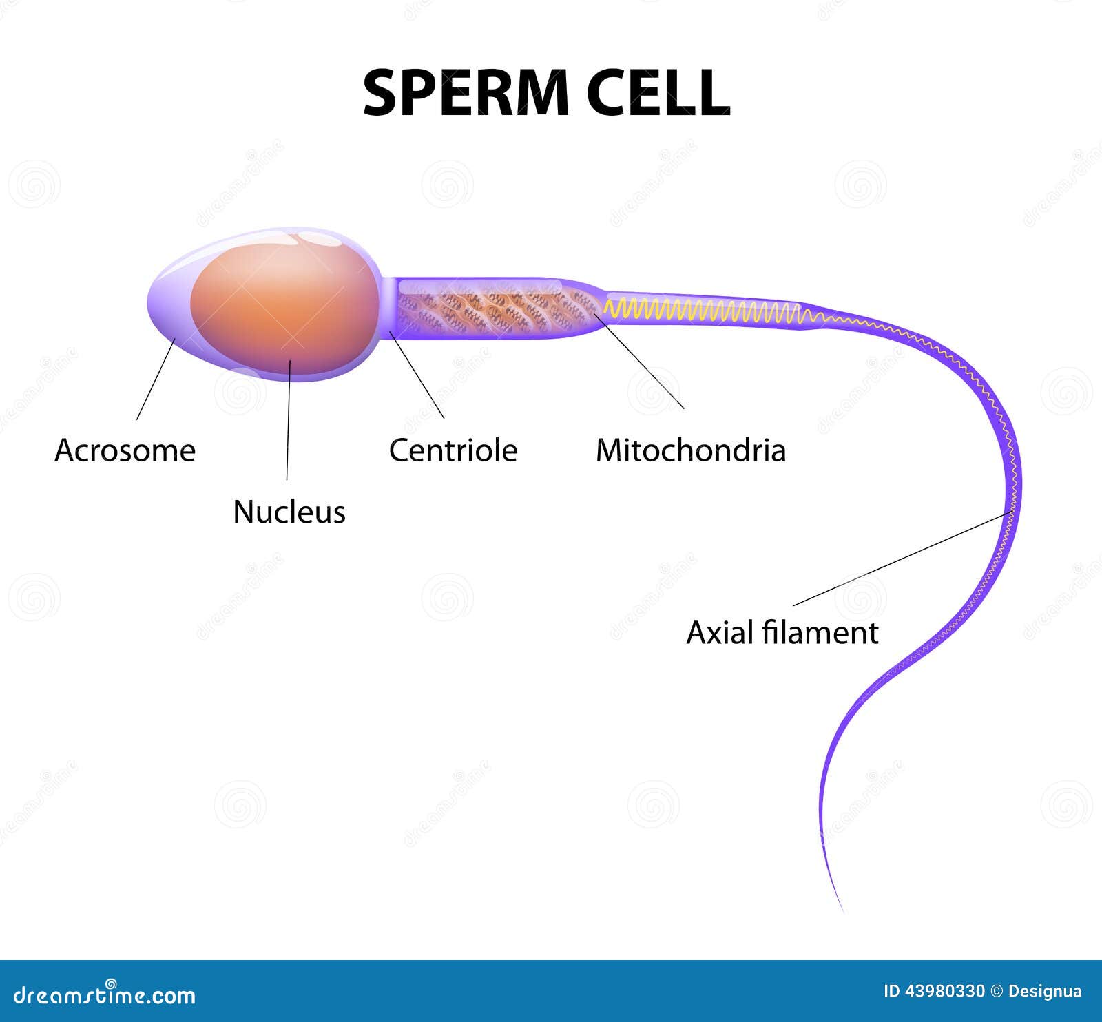
Structure of a sperm cell stock vector. Illustration of human - 43980330
Male Reproductive System: Labeled Diagram of Organs - Study.com The epididymis is a coiled tube present on each testicle that hosts sperm after they are produced. Sperm stored in the epididymis undergo further maturation, acquire motility, and reside there...

Sperm Cells for Artificial Reproduction and Germ Cell Transplantation - European Urology Supplements
quizlet.com › 515111566 › ch-8-mastering-biologych 8 mastering biology Flashcards | Quizlet Can you label the phases of the cell cycle? To review a crucial phase of the cell cycle, watch this BioFlix animation: Mitosis. Part A - The cell cycle Drag the pink labels onto the pink targets to identify the two main phases of the cell cycle. Then drag the blue labels onto the blue targets to identify the key stages that occur during those ...
Reproductive System Worksheet Answers - WikiEducator
What's the Function of a Sperm Cell? - Definition & Structure A spermatozoon, in plural spermatozoa, or sperm cell is the male reproductive cell that is expelled along with the seminal fluid or semen when a man ejaculates. In humans, spermatozoa determine the gender of the baby-to-be, which means that they can carry either the X or the Y chromosome.

Line Diagram Of A Sperm - ClipArt Best
Draw the diagram of human sperm and label its parts. Write few lines ... Draw the diagram of human sperm and label its parts. Write few lines about it. Medium Solution Verified by Toppr The sperm cells are the haploid gametes which are produced in the male. There are different parts of the sperm cell. (a) Acrosome: This structure contains enzymes used for penetrating the female egg.

BIOLOGY CST PracticeReleased California State BiologyTest Questions. © California Department of ...
Structure of Human Sperm: Check Types of Sperm - Embibe Explain the Structure of Human Sperm with Labelled Diagram Fig: Structure of a sperm cell Learn Exam Concepts on Embibe What is the Structure of Sperm? Human sperm is a microscopic structure whose shape is like a tadpole. It has flagella which make it motile. Its diameter is \ (2 - 5 {\rm { \mu m}},\) and its length is \ (60 {\rm { \mu m}}.\)

Organelles of sperm
Sperm Cell Labeled Diagram Stock Vector (Royalty Free) 200461103 ... Frequently used Trendsetter We're seeing significant engagement with this asset. Item ID: 200461103 Sperm Cell Labeled Diagram Formats EPS 6733 × 3563 pixels • 22.4 × 11.9 in • DPI 300 • JPG Contributor j joshya Similar images See all Assets from the same collection See all Similar video clips

Reproduction flashcards | Quizlet
cdn.savemyexams.co.uk › uploads › 2022/03/0970_s19Cambridge Assessment International Education ... - Save My Exams 29 A human zygote is a diploid cell. Which statement about human diploid cells is correct? A They do not have a nucleus. B They fuse to form gametes. C The nucleus contains a single set of chromosomes. D The nucleus contains two sets of chromosomes. 30 Which feature allows the sperm to dissolve the jelly coating of the egg cell? A acrosome B ...
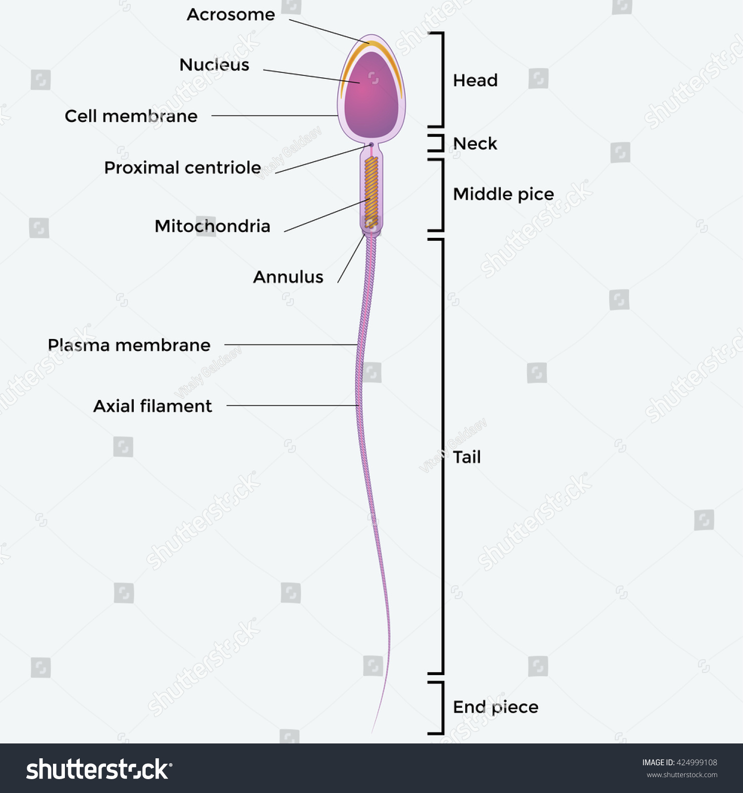
Illustartion Showing Structure Sperm Cell Stock Vector 424999108 - Shutterstock
Sperm Cell - The Definitive Guide | Biology Dictionary A sperm cell or spermatozoon is a gamete (sex cell) produced in the male reproductive tract. It is a motile cell with a single aim - to fertilize a female egg. Each sperm cell contains the entire genome of the male that produces it. In combination with the female genome contained within the egg, a zygote is formed - a single totipotent stem ...

Brenda's A & P Eportfolio: Objective 71 & 72: How sperm move and evolutionary fitness
Sperm Cell Function & Structure | What is a Sperm? The three anatomical features of a sperm cell are: 1) the head, 2) the mid-piece and lastly 3) the tail or the flagellum. The head contains the genetic material and acrosome. The mid piece contains...
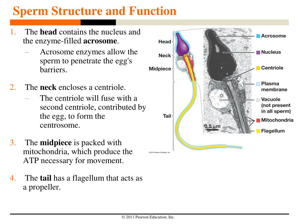
PPT - Sperm Structure and Function PowerPoint Presentation, free download - ID:4130804
Pathway of sperm: MedlinePlus Medical Encyclopedia Image The testes are where sperm are manufactured in the scrotum. The epididymis is a tortuously coiled structure topping the testis, and it receives immature sperm from the testis and stores it for several days. When ejaculation occurs, sperm is forcefully expelled from the tail of the epididymis into the deferent duct. Sperm then travels through ...

Sperm Cell Diagram Labeled - ClipArt Best
Draw a diagram of the microscopic structure of human sperm. Label the ... The above diagram is of the sperm cell. (a) Acrosome: It contains enzymes used for penetrating the female egg. (b) Nucleus: Contains the genetic material that the sperm has to pass on, a haploid genome because it contains only one copy of each chromosome.

What is the structure of a mature human sperm cell? - Lifeeasy Biology: Questions and Answers
Draw a Diagram of the Microscopic Structure of Human Sperm. Label the ... Answer in Brief Diagram Draw a diagram of the microscopic structure of human sperm. Label the following parts in it and write their functions (a) Acrosome (b) Nucleus (c) Middle piece Draw a diagram of a mature human sperm. Label any three parts and write their functions. Explain the structure of human sperm with labelled diagram.

Second Questions
Sperm Diagram Stock Illustrations - 415 Sperm Diagram Stock ... Download 415 Sperm Diagram Stock Illustrations, Vectors & Clipart for FREE or amazingly low rates! ... Ocean depth zones infographic, vector illustration labeled diagram. Oceanography science educational graphic information. Depth at which sperm whales live and. ... Blue sperm cell vector illustration. 3d fertilisation isolated.
Post a Comment for "38 sperm cell diagram with labels"